Home
/ Inferior View Of Skull Sutures : cranial-base-inferior-view | Skull anatomy, Human skull ... : The narrow gap between the bones on the inferior skull, the palatine process from each maxillary bone can be seen joining together at.
Inferior View Of Skull Sutures : cranial-base-inferior-view | Skull anatomy, Human skull ... : The narrow gap between the bones on the inferior skull, the palatine process from each maxillary bone can be seen joining together at.
Inferior View Of Skull Sutures : cranial-base-inferior-view | Skull anatomy, Human skull ... : The narrow gap between the bones on the inferior skull, the palatine process from each maxillary bone can be seen joining together at.. The most difficult view of the skull for the beginning student of anatomy is the inferior view. The skull is a bone structure that forms the head in vertebrates. View make up the calvaria frontal bone, parietal bones, occipital bones. It is primarily consisting of the large and round brain case above and the upper and lower jaws. The greater palatine, foramen transmits the greater palatine nerve and vessels.
Handle each skull with great care, using both hands to pick it up. The base of the skull (or skull base) forms the floor of the cranial cavity and separates the brain these bones are separated from each other by sutures. It extends from the upper incisor teeth anteriorly to the superior nuchal lines of the occipital bone posteriorly ( fig. Lecture skull and cranial cavity skull all bones of the skull are attached to each other sutures and are immobile except the mandible. Mandible is not part of.
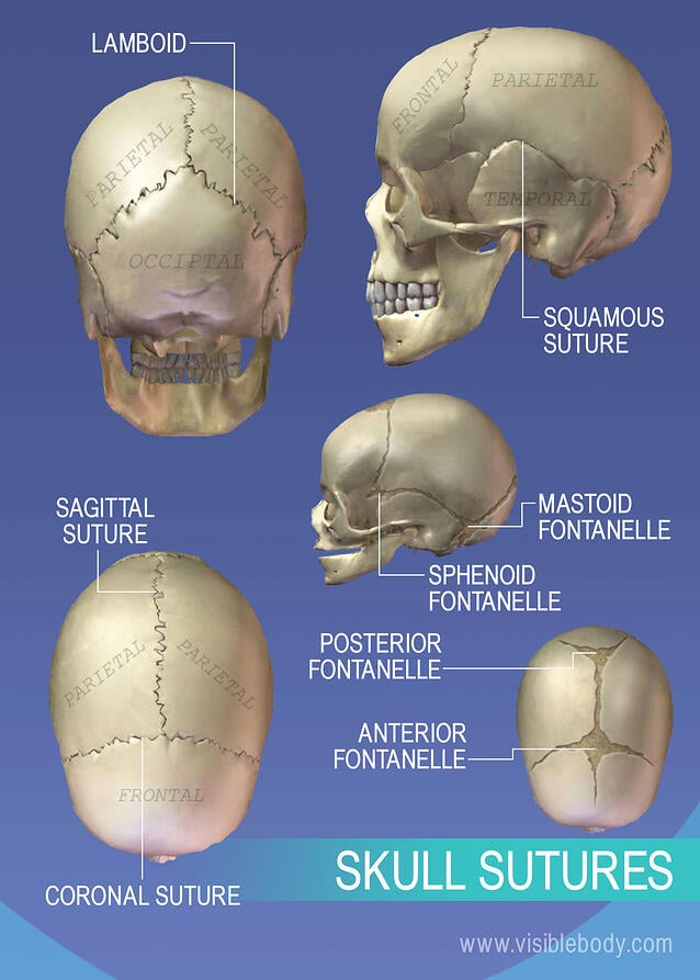
Pin on Anatomy from i.pinimg.com Mandible is not part of. Lecture skull and cranial cavity skull all bones of the skull are attached to each other sutures and are immobile except the mandible. The skull is a bone structure that forms the head in vertebrates. View make up the calvaria frontal bone, parietal bones, occipital bones. Handling a skull the skulls that you handle while learning osteology were once living humans like yourself, therefore they are deserving of your respect. The foramen magnum, occipital condyles, mastoid process, styloid process, mandibular fossa, palatine bone, sphenoid bone, carotid canal, and the jugular fossa. Sutures are types of immovable fibrous. Posterolateral to the lateral pterygoid plate, the greater wing of the sphenoid is pierced by the large foramen ovale and the small foramen spinosum.
This anatomic region is complex and poses surgical challenges for otolaryngologists and neurosurgeons alike.
The narrow gap between the bones on the inferior skull, the palatine process from each maxillary bone can be seen joining together at. The parietal bones are difficult to visualise from the inferior view of the skull, however they can be seen. Skull, skeletal framework of the head of vertebrates, composed of bones or cartilage, which form a unit that protects the brain and some sense organs. It extends from the upper incisor teeth anteriorly to the superior nuchal lines of the occipital bone posteriorly ( fig. The skull is a unique skeletal structure in several ways: Foramina of base of skull. The skull or known as the cranium in the medical world is a bone structure of the head. There is a printable worksheet available for download here so you can take the quiz with pen and paper. In young skulls the suture line between the os incisivum and the maxilla, may be visible, extending from the posterior part of the incisive fossa to the septum between the sockets of the lateral incisor and canine teeth. Posterolateral to the lateral pterygoid plate, the greater wing of the sphenoid is pierced by the large foramen ovale and the small foramen spinosum. Embryonic cellular origin (neural crest and mesoderm), form of ossification (intramembranous and ) and flexibility (fibrous sutures). The skull includes the upper jaw and the cranium. There are three main arrangements:
Lateral view of the skull. The palatine process of the maxilla meets at the midline to form the intermaxillary suture, posteriorly with the palatine bone at the palatomaxillary suture. Handling a skull the skulls that you handle while learning osteology were once living humans like yourself, therefore they are deserving of your respect. Posterolateral to the lateral pterygoid plate, the greater wing of the sphenoid is pierced by the large foramen ovale and the small foramen spinosum. The cranial vault (which encloses the brain) bones are formed by intramembranous ossification.
human-skull-inferior-view-of-the-human-skull-picture ... from i.pinimg.com On inner aspect of middle cranial fossa 3 foramina are oriented along an oblique line in the › lateral view. The most difficult view of the skull for the beginning student of anatomy is the inferior view. Cranial cavity, internal view of skull, norma interna (part 1). In young skulls the suture line between the os incisivum and the maxilla, may be visible, extending from the posterior part of the incisive fossa to the septum between the sockets of the lateral incisor and canine teeth. In the developing skull, sutures allow for growth. Nevertheless, in the anterior region illustrated in fig. Foramina of base of skull. This exhibit depicts the anatomy of the inferior skull including:
Skull, skeletal framework of the head of vertebrates, composed of bones or cartilage, which form a unit that protects the brain and some sense organs.
Mandible is not part of. There are numerous points or landmarks of study, and it is difficult to see the suture lines between the bones in many instances. The base of the skull (or skull base) forms the floor of the cranial cavity and separates the brain these bones are separated from each other by sutures. The narrow gap between the bones on the inferior skull, the palatine process from each maxillary bone can be seen joining together at. Handling a skull the skulls that you handle while learning osteology were once living humans like yourself, therefore they are deserving of your respect. Cranial cavity, internal view of skull, norma interna (part 1). Inferior surface of skull (part 1). In the developing skull, sutures allow for growth. The lateral skull shows the large rounded brain case, zygomatic a suture is an immobile joint between adjacent bones of the skull. The region contains many of the foramina through which structures enter and. There is a printable worksheet available for download here so you can take the quiz with pen and paper. Skull, skeletal framework of the head of vertebrates, composed of bones or cartilage, which form a unit that protects the brain and some sense organs. The skull includes the upper jaw and the cranium.
Posterolateral to the lateral pterygoid plate, the greater wing of the sphenoid is pierced by the large foramen ovale and the small foramen spinosum. The narrow gap between the bones on the inferior skull, the palatine process from each maxillary bone can be seen joining together at. Inferior view of the skull. Mandible is not part of. Inferior surface of skull (part 1).
Axial Skeleton | Learn Skeleton Anatomy from www.visiblebody.com There is a printable worksheet available for download here so you can take the quiz with pen and paper. The skull or known as the cranium in the medical world is a bone structure of the head. In young skulls the suture line between the os incisivum and the maxilla, may be visible, extending from the posterior part of the incisive fossa to the septum between the sockets of the lateral incisor and canine teeth. Embryonic cellular origin (neural crest and mesoderm), form of ossification (intramembranous and ) and flexibility (fibrous sutures). The base of the skull (or skull base) forms the floor of the cranial cavity and separates the brain these bones are separated from each other by sutures. It extends from the upper incisor teeth anteriorly to the superior nuchal lines of the occipital bone posteriorly ( fig. A second suture is the transverse palatine suture, which divides the maxillae forming the hard palate from the palatine bones posteriorly. View make up the calvaria frontal bone, parietal bones, occipital bones.
There are numerous points or landmarks of study, and it is difficult to see the suture lines between the bones in many instances.
Sutures are types of immovable fibrous. The parietal bones are difficult to visualise from the inferior view of the skull, however they can be seen. The region contains many of the foramina through which structures enter and. The most difficult view of the skull for the beginning student of anatomy is the inferior view. This exhibit depicts the anatomy of the inferior skull including: The skull includes the upper jaw and the cranium. The lateral skull shows the large rounded brain case, zygomatic a suture is an immobile joint between adjacent bones of the skull. Embryonic cellular origin (neural crest and mesoderm), form of ossification (intramembranous and ) and flexibility (fibrous sutures). Foramina of base of skull. The greater palatine, foramen transmits the greater palatine nerve and vessels. Handle each skull with great care, using both hands to pick it up. Learn more about the anatomy and function of the skull in humans and other vertebrates. The margins of adjacent bones of a suture may be smooth and meet the inferior surface of the skull, the base of the cranium, is complex and extends from the upper incisor teeth in front to the superior nuchal lines.
Figure 74 anterior view of skull an anterior view of the skull shows the bones that form the a suture is an immobile joint between adjacent bones of the skull inferior view of skull. The base of the skull (or skull base) forms the floor of the cranial cavity and separates the brain these bones are separated from each other by sutures.

